
FIGURE 1.
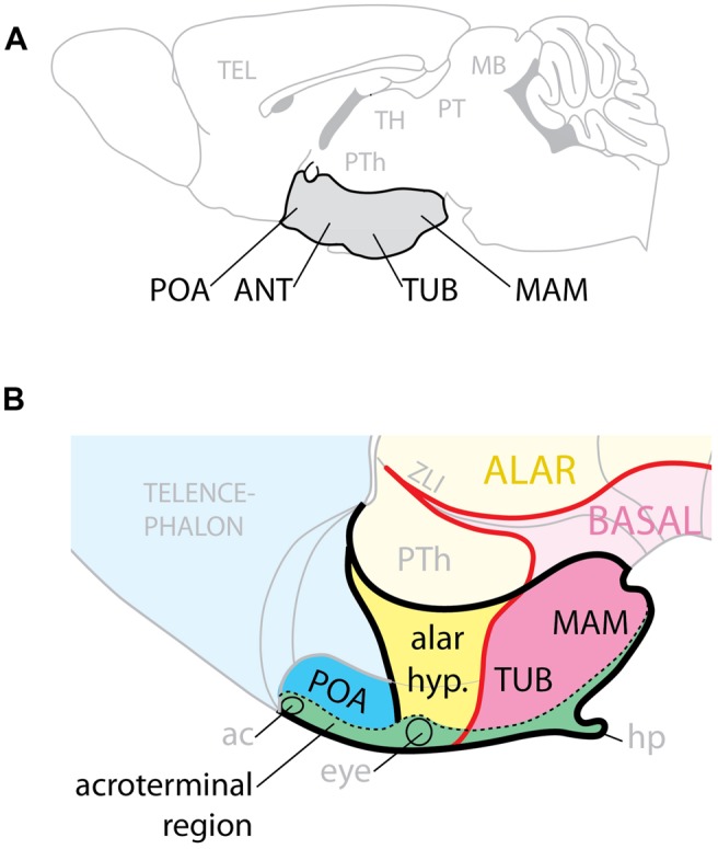
Hypothalamic regions. (A) Conventional representation of the hypothalamus (gray) as ventral region with four rostro-caudal regions, POA, preoptic; ANT, anterior; TUB, tuberal; MAM, mamillary. (B) Model of the hypothalamus considering Shh expression (pink) as basal (ventral) marker. The POA is part of the telencephalon; the alar hypothalamus (yellow) corresponds to the anterior region; the tuberal and mamillary regions are not “caudal” but basal (ventral). ac, anterior commissure; hp, hypophysis; PTh, prethalamus; ZLI, zona limitans.