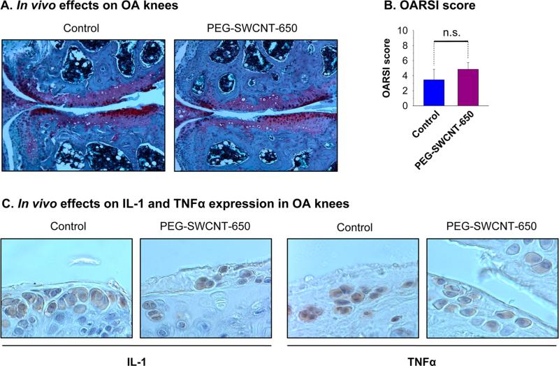
Figure 4.
In Vivo effects of IA-injected PEG-SWCNTs. Two groups (N = 4) of 2-month-old female B6 mice received DMM surgery and were IA-injected with either 5 μg of IA-PEG-SWCNTs in 10 μL of PBS or an equal volume of vehicle (PBS, control), in the OA knee. After 2 months, OA knees were collected, fixed, decalcified, and embedded in paraffin. Six micrometer thick frontal sections were taken at 60 μm intervals and stained with Safranin-O and counterstained with Fastgreen and Hematoxylin (A). Cartilage damage was examined on the four knee quadrants and scored using the OARSI method. There was no statistically significant cartilage worsening in PEG-SWCNT-650-treated knees respect to control knees. Statistics: nonparametric Mann–Whitney U analysis (n.s. = not significant) (B). Alternatively, sections were stained with either anti-IL-1 or anti-TNα antibodies. IL-1 and TNFR expression was examined on the four knee quadrants. There was no protein overexpression in PEGSWCNT-650-treated knees respect to control knees of OA mice (C).

