Fig 3.

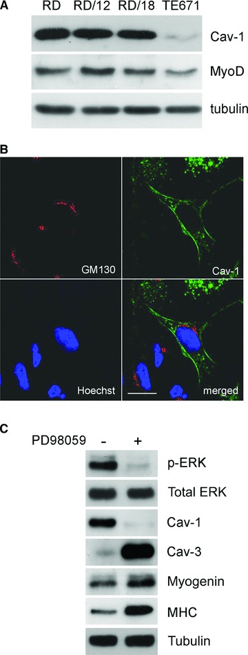
Expression of caveolins in RMS cell lines. (A) Western blot analyses showing the expression of Cav-1 in several MyoD-positive ERMS cell lines. Tubulin was used as loading control. (B) As shown by confocal microscopy analysis, Cav-1 localizes at the plasma membrane or in intracellular vesicles of embryonal RD cells. GM130 marker was employed to stain the Golgi apparatus. Bars = 100 μm. (C) Ten microliters of PD98059 administration attenuates the ERK phosphorylation in RD cells and allows the transition from proliferation to differentiation, leading to Cav-1 down-regulation and increase of myogenin, MHC and Cav-3. Tubulin was used as loading control.
