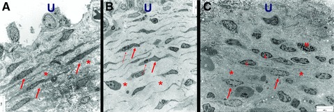
Fig 1.

Electron micrograph of ULP ICLC in control (A), NDO, (B) and BPS (C) bladders. (A) Many parallel layers of ICLC (arrows) embedded in a dense extracellular matrix (asterisk). (B) Multiple parallel layers of slender ICLC (thick arrow) often in close association with lymphocytes (thin arrow). Note the large intercellular space (asterisk) filled with less dense intercellular matrix components. (C) Fragmented layers of ICLC (arrows). Note the presence of lymphocytes (l) and mast cells (m). Dense extracellular matrix components are obvious (asterisk). Scale bars: 7 μm. U: urothelial area.
