
Fig 5.
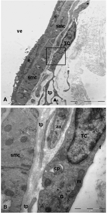
Transmission electron microscopy images of telocyte in close apposition to wall of venule. (A) A typical example of telocyte: long, thin and moniliform processes with dichotomous pattern (arrowhead) of branching. Black square illustrating a higher magnification of telocyte from A. (B) The perinuclear cytoplasm contains well-developed Golgi apparatus (G), mitochondria (m), numerous polyribosomes (asterisk) and intermediate filaments (f). Note the deep invagination (cilium-pit) corresponding to primary cilium. smc: smooth muscle cells; tp: telopodes; ax: axon; TC: telocyte; e: endothelial cell; cp: cilium-pit; pd: podom; ve: venule.