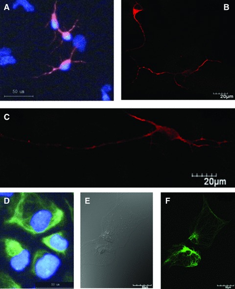
Fig 3.

Morphologies and biomarker-specific staining of differentiated neurons and astrocytes. The cell showed positive to the staining of MAP-2 and the nuclear straining with Hoechst 33342 (A), and formed clear neurites (B) and clear neural axons and dendrites in the intermediate phase of differentiation from NSCs to neurons (C). NSCs-differentiated astrocytes were detected in the microscopy with (D) and without GFAP staining (E). Some showed the matured shape of astrocytes with rich and strong GFAP staining (F).
