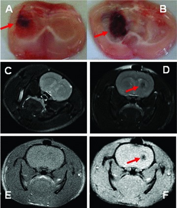
Fig 4.

The formation of intracerebral haematoma was detectable at 6 hrs (A) and reached the maximal 3 days after the injection of collagenase (B). The examination of the nuclear magnetic resonance imaging showed the uniform of the density in normal brain at AX FSE T2W scan (C), the round lesion with low density in ICH brain (D) and the clearer lesion at high resolution of GRE T2WI (E), as compared with FSE T1WI resolution (F), 3 days after the induction of ICH. The lesions or abnormalities were pointed with red arrow.
