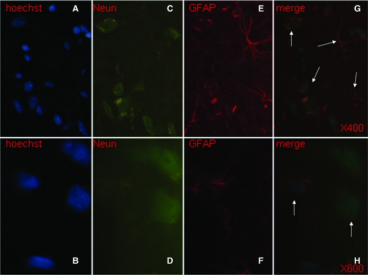
Fig 7.

Transplanted foetal NSCs in the brain showed positive to the single staining with Hoechst (blue, × 400 and × 600, A and B), NeuN (green, × 400 and × 600, C and D) and GFAP (red, × 400 and × 600, E and F), and to triple staining of Hoechst, NeuN and GFAP ( × 400 and × 600, G and H). The differentiated neurons had double staining of Hoechst and NeuN, whereas the astrocytes had Hoechst and GFAP, as pointed with arrows.
