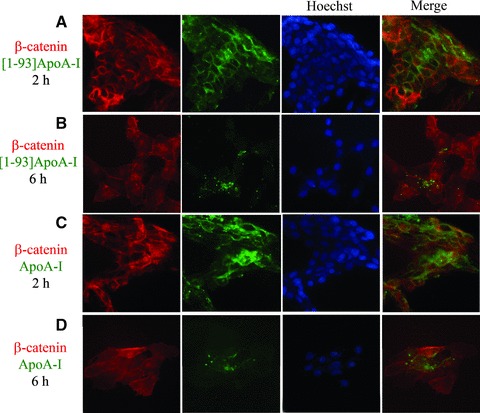
Fig 2.

Endocytosis of [1–93]ApoA-I and full-length ApoA-I in H9c2 cells. Cells were grown on cover slips, incubated 2 hrs (A) or 6 hrs (B) with 3 μM FITC-[1–93]ApoA-I (green) and immunofluorescently stained for β-catenin (red). (C) and (D), cells incubated 2 or 6 hrs, respectively, with 1 μM FITC-ApoA-I (green) and immunostained for β-catenin (red). Nuclei were stained with Hoechst (blue). Cells were analysed by epifluorescence microscopy.
