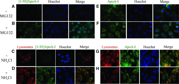
Fig 6.

Analysis of the degradation pathway of [1–93]ApoA-I and ApoA-I in H9c2 cells by epifluorescence microscopy. (A)–(D) [1–93]ApoA-I degradation. Cells were incubated 24 hrs at 37°C with 3 μM FITC-[1–93]ApoA-I in the absence (A, C) or in the presence of MG132 (2.5 μM) (B), or ammonium chloride (100 μM) (D). (E)–(H), ApoA-I degradation. Cells were incubated for 24 hrs at 37°C with 1 μM FITC-ApoA-I, in the absence (E and G) or in the presence of MG132 (F), or ammonium chloride (H). Lysosomes were stained with LysoTracker red. Nuclei were stained with Hoechst (blue).
