
Fig 1.
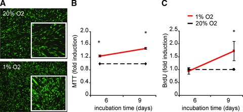
Hypoxia increases the proliferation of hCMPCs. hCMPCs were cultured under normoxic and hypoxic conditions for the indicated time intervals. An increase in the number of cells under hypoxia was visualized by staining with fluorescein di-acetate (A). Viability and cell cycle progression was quantified by MTT (B) and BrdU (C), respectively. Both assays show an increased proliferation under hypoxia. Values were normalized to normoxia of the same time-point and one representative experiment of seven is shown. *Significant difference between normoxia and hypoxia at the same time-point.