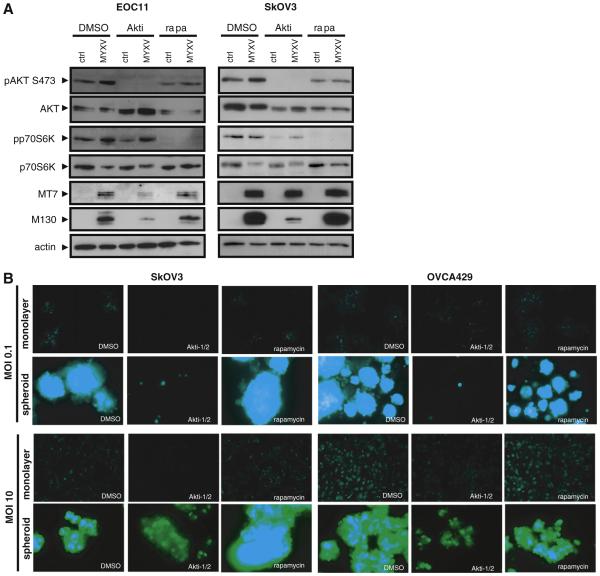
Fig. 4.
AKT activity is required for efficient MYXV infection of EOC cells and spheroids. (A) Western blotting was performed to determine expression of phosphorylated AKT kinase and phosphorylated p70 S6 kinase at 24 h post-MYXV infection of primary EOC11 cells and SkOV3 cells grown in monolayer culture and treated with the AKT inhibitor Akti-1/2 (5 μM), the mTOR inhibitor rapamycin (20 nM), or DMSO vehicle control. Phosphorylated AKT was completely absent in Akti-1/2 treated cells and this markedly decreased MYXV infection as determined by M-T7 and M130 expression. MYXV infection increased phospho-p70S6 kinase, which is unaltered by Akti-1/2 treatment. Rapamycin treatment did not affect MYXV infection, but was effective at reducing phospho-p70 S6 kinase levels to below detection. Actin served as a loading control. (B) SkOV3 and OVCA429 monolayer cells and spheroids were infected with MYXV (MOI of 0.1 and 10) treated with the AKT inhibitor Akti-1/2 (5 μM), the mTOR inhibitor rapamycin (20 nM), or DMSO vehicle control. Images were captured by fluorescence microscopy at 96 h post-infection for MOI of 0.1 and 24 h post-infection at an MOI of 10. Treatment with Akti-1/2 decreased the efficiency of MYXV infection in both monolayer and spheroids when compared to rapamycin treatment and DMSO controls as determined by intensity and degree of fluorescence.

