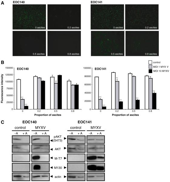
Fig. 5.
Ascites may reduce the efficiency of MYXV infection and oncolysis of primary EOC cells. Two independent primary EOC samples (EOC140 and EOC141) were infected with MYXV at an MOI of 1 and 10, or UV-inactivated control virus, while in the presence of clarified ascites (0, 20, 50 or 80% diluted in complete media) which was matched to the same patient. (A) Marked reduction of MYXV infection (MOI 10) was observed in EOC140 at all ascites concentrations tested when compared with complete media alone control (0 as- cites); ascites had little to no observable effect on infection in EOC141. (B) MYXV-mediated oncolysis of EOC140 cells was inhibited by the presence of ascites at all concentrations tested for both MYXV doses when compared with complete media alone control, as determined by alamarBlue® assay after 3 days of treatment. Reduction in EOC141 cell viability by MYXV oncolysis was largely unaffected at an MOI of 10, whereas oncolysis at the lower dose (MOI 1) was inhibited by increasing concentration of ascites. Note that ascites fluid alone (control, UV-inactivated virus) did not affect the overall viability of EOC cells over the 3-day treatment period at all concentrations tested. (C) Western blot analysis was per- formed using lysates from EOC140 and EOC141 cells to determine phospho-AKT (Ser473) and total AKT protein expression levels in the presence (+ A) or absence ( A) of ascites (20% in complete media) after 24-hour treatment and concomitant infection with MYXV at an MOI of 1. Detection of M-T7 and M130 proteins confirmed MYXV infection of EOC cells in the absence of ascites, whereas M-T7 and M130 were only present in EOC141 cells in the presence of ascites, which correlates with cell viability data.

