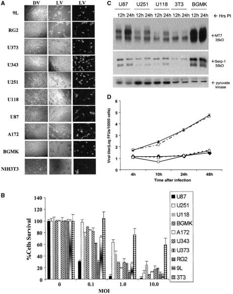
Figure 1.
Myxoma virus infects and kills most glioma cells in vitro. A, glioma cells were infected with live vMyxgfp (MOI = 10, left and center column) or dead virus (dead myxoma virus, i.e., UV-inactivated vMyxgfp, right column). Cytopathic effect and green fluorescent protein expression were evident in all live vMyxgfp–infected cell lines, but not in the dead myxoma virus-infected cell lines, 72 hours after infection (magnification, ×100). B, cells infected with different MOIs of vMyxgfp were evaluated for viability by MTT assay at 72 hours postinfection. U87 and RG2 cell lines were very susceptible. The U118 cell line was much less susceptible and a significant proportion of cells were killed only at 96 hours and an MOI of 10 (data not shown). BGMK and NIH 3T3 were used as positive and negative controls, respectively. C, myxoma viral protein synthesis was evident after virus infection. An increase in both early (M-T7) and late (Serp-1, an indicator of virus replication) viral gene products was detected in glioma lines and BGMK by Western blot analysis, 12 to 24 hours after myxoma virus infection (MOI = 5). Pyruvate kinase was used as a protein loading control. D, myxoma virus replicates within glioma cells. Cells were infected with vMyxgfp (MOI = 0.1) followed by collection of cell lysates at the indicated times after infection. Viral titers were determined by plaque titration on BGMK cells. Viral titers increased in cell lysates of all glioma cell lines although only minimally in U118 and U251. —◆–, U118; —○—, U87; —▲—, BGMK; —□—, U251; —△—, 3T3.

