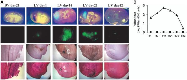
Figure 4.

Myxoma virus infection is selective, productive, and produces a long-lived infection in human gliomas in vivo. A, vMyxgfp expression was limited to the tumor and was not found in any normal brain areas under bright- and dark-field imaging (first and second rows, respectively). Fourth row, low-magnification images of H&E-stained sections. U87 allografts were established followed by intratumoral vMyxgfp inoculation. Animals were sacrificed at different times postinfection and tumor-containing brain regions and regions of the brain without visible tumor were examined using immunohistochemistry for myxoma virus antigens (third row) and fluorescent imaging of green fluorescent protein expression (second row). Green fluorescent protein expression and viral antigens were visualized only at the site of viral inoculation in the live vMyxgfp–treated animals. No evidence of virus replication was detected in the dead myxoma virus–treated animals. B, quantification of intratumoral virus replication in virus inoculation side (—▲—) and contralateral side (—■—) for both live vMyxgfp– and dead myxoma virus–treated animals. Virus titers (log PFUs/10,000 cells) at days 1, 7, 14, 21, and 42 postinfection [points, mean (n = 3/time point); bars, SD]. Virus titers increased by >1 log and peaked 14 days after inoculation in virus inoculation (tumor) side. Both dead myxoma virus–treated tumors (not shown) and normal brain regions (contralateral) had no viable virus recovered. Viral cultures were done on BGMK cells by plaque assay.
