Figure 1.

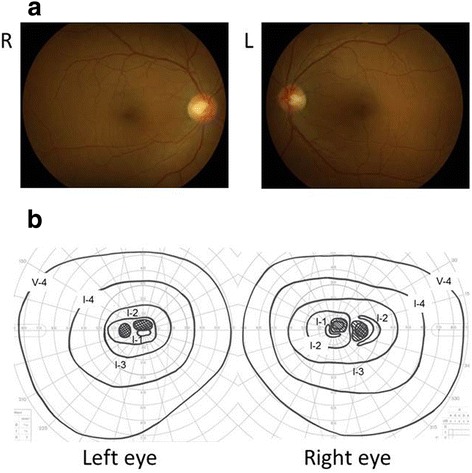
Fundus photograph (a) and Goldmann perimetry (b). Fundus appearances looked normal and central scotomas were noted in both eyes at the initial visit.

Fundus photograph (a) and Goldmann perimetry (b). Fundus appearances looked normal and central scotomas were noted in both eyes at the initial visit.