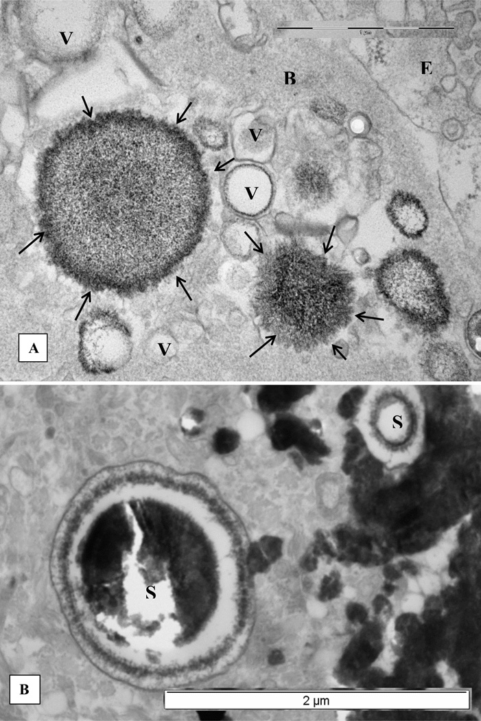
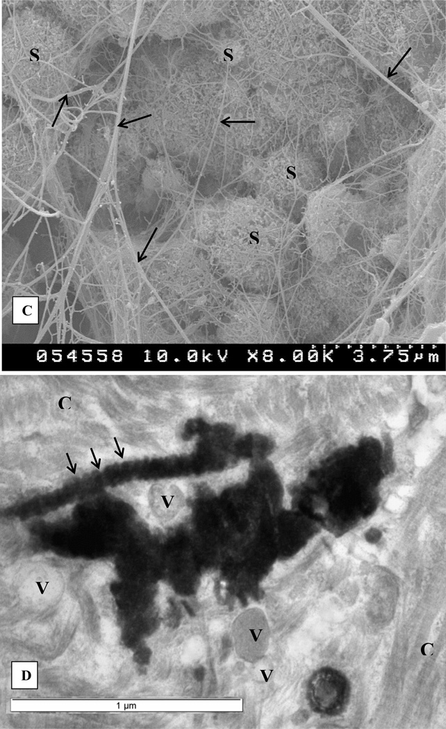
Figure 1.
Electron microscopic examination of renal interstitial deposits of the CaP (For details please refer to [26]. A. Membrane bound vesicles associated with the basement membrane (B) of an epithelial cell (E). Some vesicles appear empty (V) while others contain electron dense material with needle shaped crystals of apatite on their periphery (arrows). B. Edge of a dense calcium phosphate deposit showing two large laminated spherical (S) bodies of apatite crystals. C, collagen. C. Calcium phosphate deposits as seen by scanning electron microscopy. Spherical apatite bodies (S) of different sizes are aggregated together with fibrous material (arrows) of various thicknesses. Needle shaped apatite crystals are sticking out on the surface of the spherical bodies similar to what is seen in Figure 1B by transmission electron microscopy. D. Another area of the dense calcium phosphate deposit shows an elongated calcified body with distinct bands (arrows). Nearby collagen fibers (C) demonstrate similar banding. V, vesicles.


