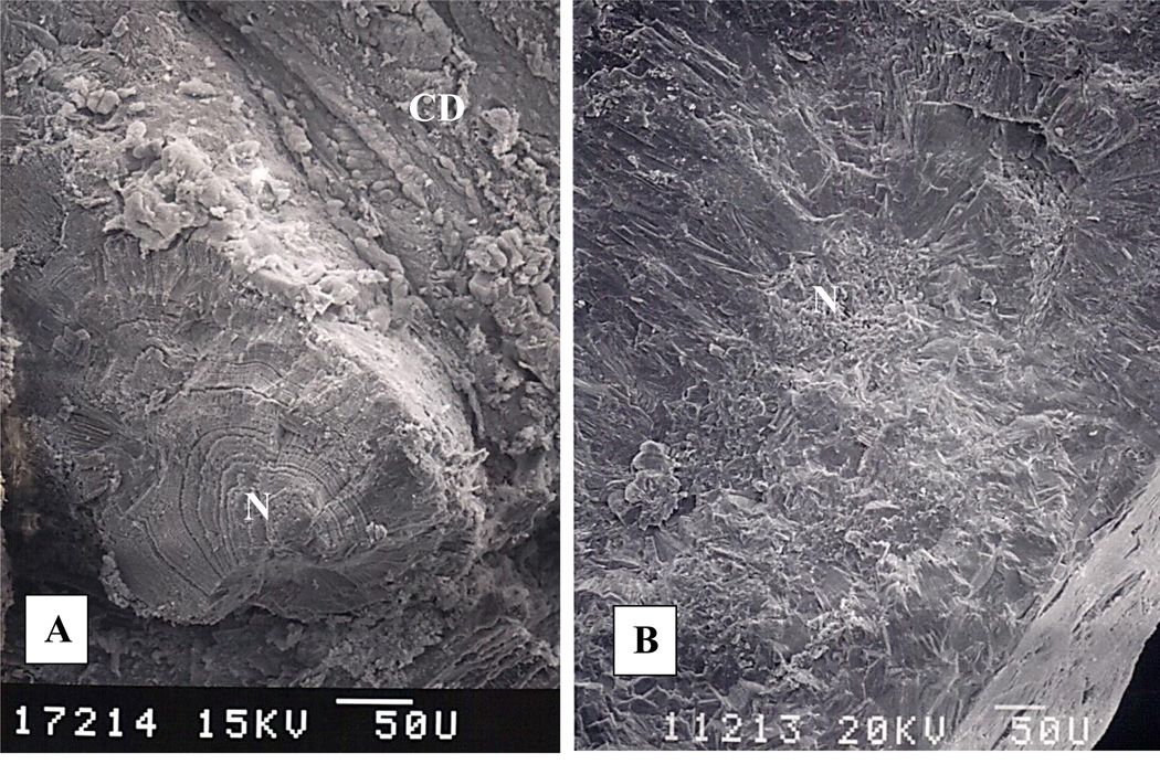
Figure 3.
Scanning electron microscopic examination of the fractured surfaces of the ductal stones. A. Stone is plugging the opening of a duct of Bellini. A nearby terminal collecting duct (CD) is on the right. Stone shows concentrically arranged layers of calcium oxalate monohydrate crystals around an acentric nucleus (N). Stone appears to have grown attached to tubular surface. B. A calcium oxalate monohydrate stone which was found free in the collecting duct. Crystals are arranged in concentric layers around a central nucleus (N).

