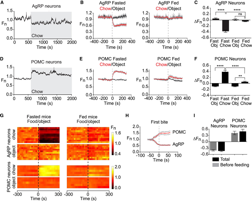
Figure 3. Sensory detection of food rapidly regulates AgRP and POMC neurons.
(A and D) Recordings from fasted mice expressing GCaMP6s in AgRP or POMC neurons presented with a pellet of chow (gray). (B and E) Plots of calcium signals from AgRP and POMC neurons aligned to the time of presentation of a pellet of chow (red) or inedible object (black). Mice were either subjected to an overnight fast (left) or fed ad libitum (right) prior to experiment. Gray indicates standard error (AgRP, n=10; POMC, n=5). (C and F) Quantification of fluorescence changes 5 min after event, as indicated. (G) Peri-event plots aligned to the time of event. Each row is a single trial of a different mouse. (H) Calcium signals aligned to the initiation of feeding for AgRP and POMC neurons. (I) Quantification of change in fluorescence occurs before feeding is initiated versus the total change in the trial. * p<0.05. ** p<0.01,*** p<0.001,**** p<0.0001.

