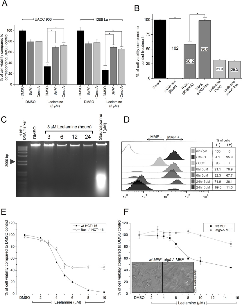
Figure 2. Lysosomotropic property of leelamine induced caspase-independent cell death leading to disruption of ΔΨm.
A, viability of melanoma cells treated with leelamine in the absence or presence of V-ATPase inhibitors BafA1 or Conc-A. B, caspase dependence of leelamine-mediated cell death measured through treatment of melanoma cells with leelamine in the absence or presence of pan-caspase inhibitor, z-VAD-fmk. C, DNA laddering assay showing absence of DNA fragmentation following leelamine treatment. Staurosporine was used as a positive control for apoptosis-mediated DNA fragmentation. D, histogram showing ΔΨm following leelamine or FCCP (positive control) treatment. E, viability of wild-type or BAX-knockout HCT116 cells after 24 hours of treatment with increasing concentrations of leelamine. F, viability of wild-type or atg5-knockout MEF cells after 24 hours of treatment with increasing concentrations of leelamine. Images showing that leelamine did not cause vacuolization of ATG5-knockout MEF cells. (*, P < 0.05, t test.)

