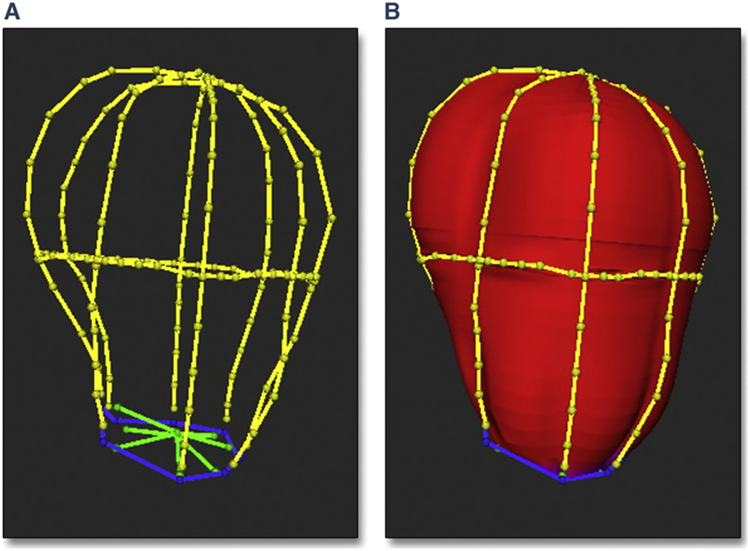
Figure 1. Endocardial Borders Conforming to Delineated Borders.
In this example, the endocardial borders of the left atrium (LA) (A) and the polygonal mesh surface conform to the delineated borders (B). Borders were traced on 4 equiangular planes (0°, 45°, 90°, and 135°) passing through the long axis of the LA and 1 cross-sectional plane perpendicular to the axis at the mid-atrium. Red indicates left atrial endocardial surface; blue indicates mitral valve annulus; yellow indicates left atrial borders; and green indicates mitral valve leaflets.

