Figure 2.
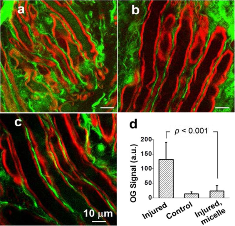
Neuroprotection from mPEG-PDLLA micelles. Calcium influx into axons. (a-c) TPEF images of OG 488 (green) and coherent anti-Stokes Raman scattering images of myelin (red) show intra-axonal free Ca2+ levels in compression-injured (a), healthy (b), and compression-injured and micelle-treated (c) spinal cords. Images were acquired 1h after compression injury. (d) Statistical analysis. Without micelle treatment, the TPEF intensity from OG inside the injured axons was 10 times greater than intact axons. The intensity was only twice that of intact axons when 0.67 mg/ml micelles were added immediately after compression injury.

