Figure 3.
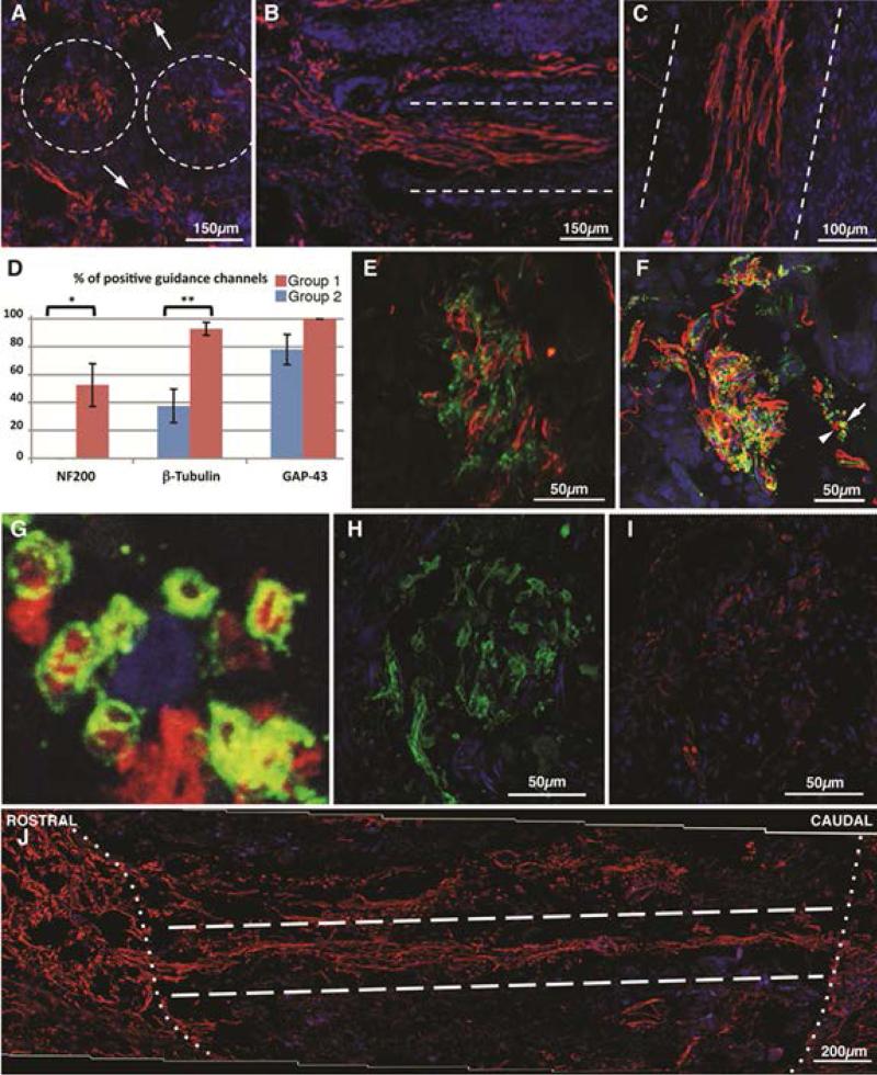
Nerve regeneration in chronically injured rat spinal cord using PLGA/PCL microchannel conduits with RADA16-1-BMHP. Dashed lines outline implanted channel walls in (A-C). In transverse sections (A), GAP-43 positive fibers were found inside and between transplanted tubes (arrows; coronal section). (B) NF200 positive fivers were seen crossing the top rostral interface of the lesion. (C) βIII-tubulin positive fasciculi stretched through the lumen of the conduits in a longitudinal spinal cord section. (D) Quantitative analysis (mean +- SEM) of the percentage of channels showing positivity for neural markers throughout the entire tube length (N=5 for group 1, N-8 for group 8). Statistical significance (*) p=0.03; (**) p = 0.003. NF200 (green) and GAP43 (red) did not colocalize inside the same fiver (E). Myelinated (green, arrow) and unmyelinated (red, arrowhead) βIII-tubulin positive fibers (F) were observed (enlargement in G). Both SMI-32 (H) and SMI-31 (I) positive fibers were detected within guidance conduits, in adjacent coronal sections. In the longitudinal reconstruction of transplanted cord in (J) (group 2), NF200 positive fibers cover the whole length (approx 2 mm) of the lesion, crossing both the rostral and the caudal channels/tissue interfaces (dotted line).

