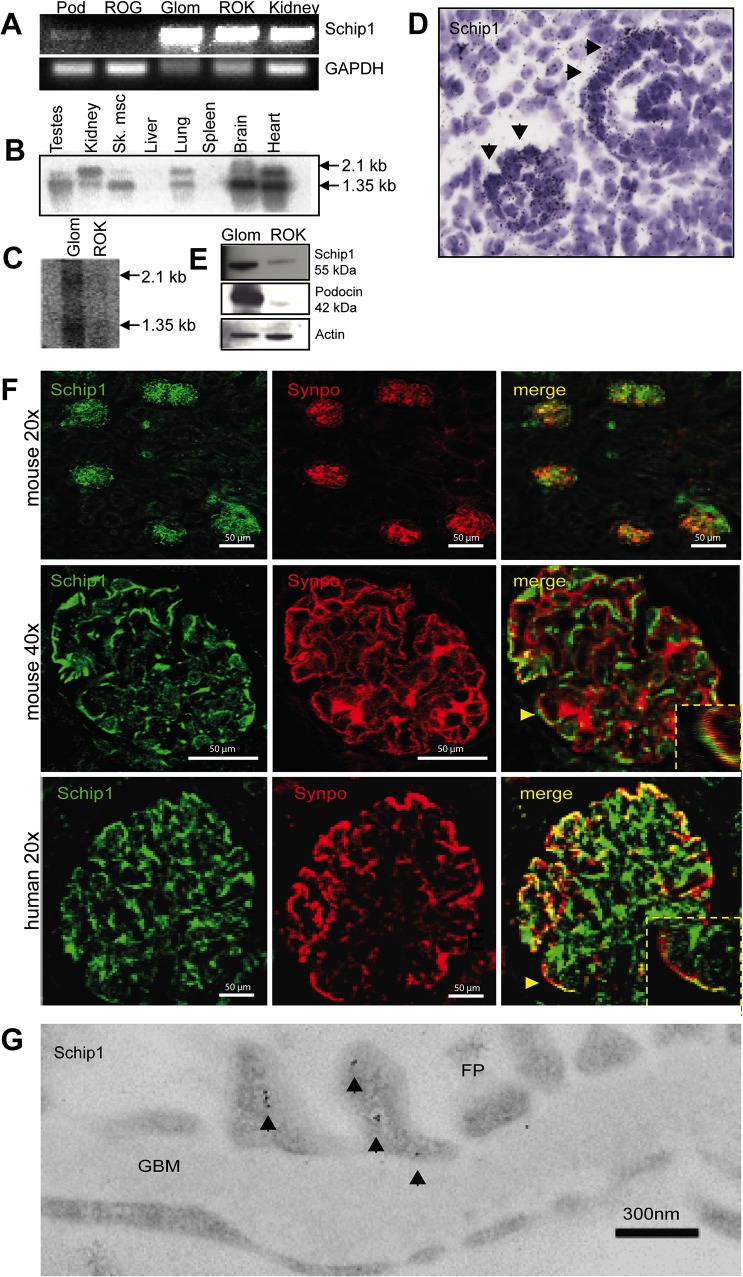
Fig 1. Schip1 is expressed by glomerular podocytes.
(A) RT-PCR shows SCHIP1 expression in both glomerulus and the kidney fraction lacking glomeruli. In the glomerulus, expression is mostly detected in FACS-sorted podocytes. As an internal control, expression levels of GAPDH were measured. Controls for markers of various fractions are presented in S3 Fig. (Pod-podocytes, ROG-rest of glomerulus, GLOM-glomerulus, ROK-rest of kidney). (B) Northern blotting on a mouse multiple tissue panel shows the presence of two SCHIP1 mRNA transcripts enriched in the brain, heart, testes and kidney tissues. (C) Northern blotting on mouse glomerulus and ROK tissue confirms stronger SCHIP1 expression in the glomerulus, and presence of two transcripts. (D) By radioactive in situ hybridization on newborn mouse kidney sections SCHIP1 mRNA is localized to developing podocytes of the capillary loop stage glomerulus. (E) By Western blotting, the mouse 55kDa Schip1 protein is detected mostly in the glomerulus. Podocin was used as a positive control for the glomerular fraction, β-actin as a loading control. (F) Immunofluorescence on mouse and human kidney sections shows Schip1 glomerulus signal that partially overlaps with a podocyte foot process marker synaptopodin (Synpo). (G) By immunoelectron microscopy, Schip1 localizes to the glomerular podocyte foot processes (FP) in human kidney sections (GBM-glomerular basement membrane).

