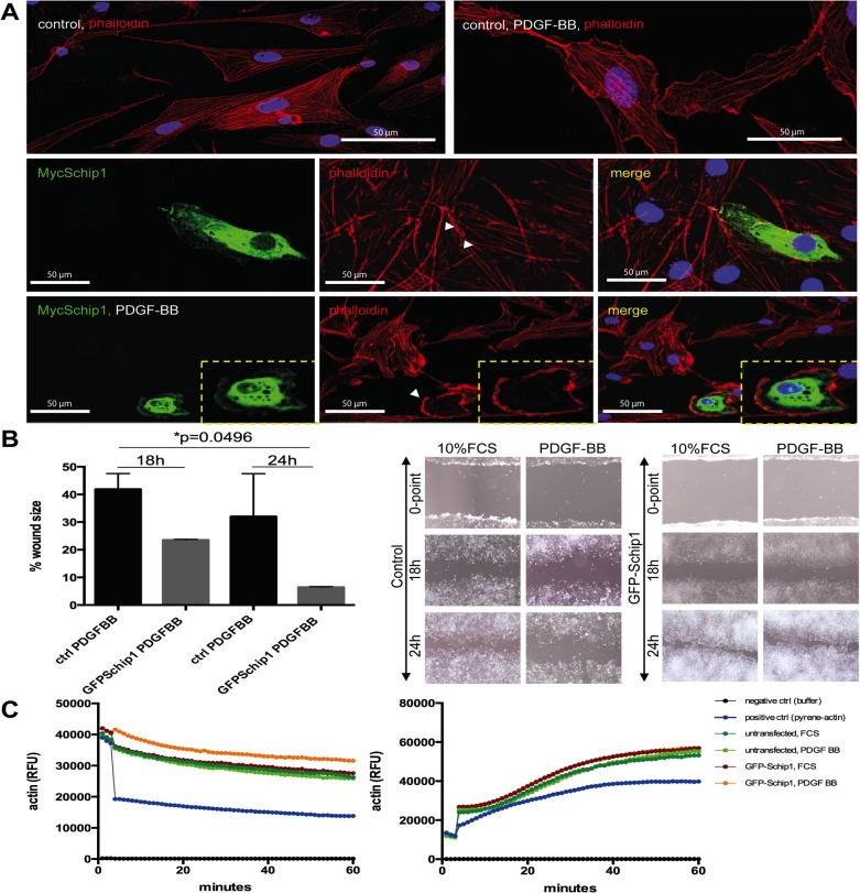
Fig 6. Schip1 overexpression promotes cortical F-actin accumulation, dissolution of stress fibers and motility in PDGF-BB-stimulated cells by attenuating actin depolymerisation.
(A) In control cells PDGF-BB treatment enhances development of lamellipodia. In Schip1-transfected cells PDGF-BB stimulation induces similar changes but also marked actin cytoskeleton rearrangement with cortical actin accumulation and dissolution of the actin stress fibers (box, zoom). Observe the neighboring non-Myc-Schip1 expressing cells, presenting with normal actin cytoskeleton and pronounced stress fibers. (B) Stable Schip1-expressing and control HEK293 cells were stimulated with 10% FCS or PDGF-BB, scratched, and left to migrate for 24 h (the wound healing assay). Schip1 transfected cells exhibit similar migration rate as controls in medium supplemented with 10% FCS, but migrate faster when induced with PDGF-BB (graph). Microscopic images of control and Schip1-expressing cell monolayers 18 and 24 h after wound scratching (left). (C) In vitro actin polymerization (right panel) and depolymerization (left panel) assays with lysates from GFP–Schip1-expressing HEK293 cells and controls show that Schip1-overexpression slows down actin depolymerization in presence of PDGFBB in comparison to cells treated with 10% FCS (p<0.0001). Results are representative of three separate experiments. RFU-relative fluorescence units.

