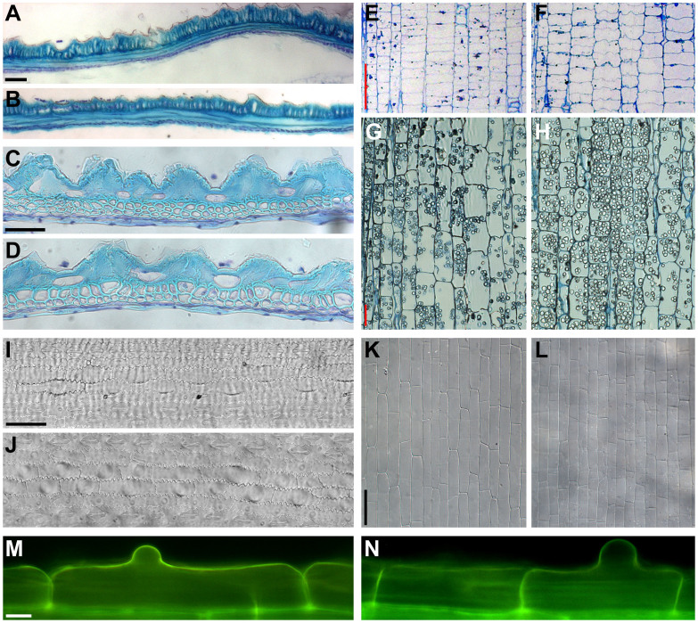
Figure 2. Cell length is reduced in sar1 glumes, internodes, leaves and roots.
(A–D) Longitudinal (A and B) and transverse (C and D) section of the lemma from mature WT (A and C) and sar1 (B and D) florets. (E–H) Longitudinal section through the intercalary meristem (E and F) and the elongation zone (G and H) of the second internode from mature WT (E and G) and sar1 (F and H) stems. (I–L) DIC observation of the leaf blade (I and J) and sheath (K and L) from mature WT (I and K) and sar1 (J and L) flag leaves. (M–N) Fluorescence microscopy of epidermal cells with visible root hair bulges in roots of WT (M) and sar1 (N). Scale bar = 50 µm in (A) for (A) and (B), in (C) for (C) and (D), in (E) for (E) and (F), in (G) for (G) and (H), in (I) for (I) and (J), in (K) for (K) and (L); bar = 10 µm in (M) for (M) and (N).

