
Fig 7. Native green gel electrophoresis of chlorophyll-protein complexes isolated from spinach thylakoid membranes treated with Al3+ at various concentrations.
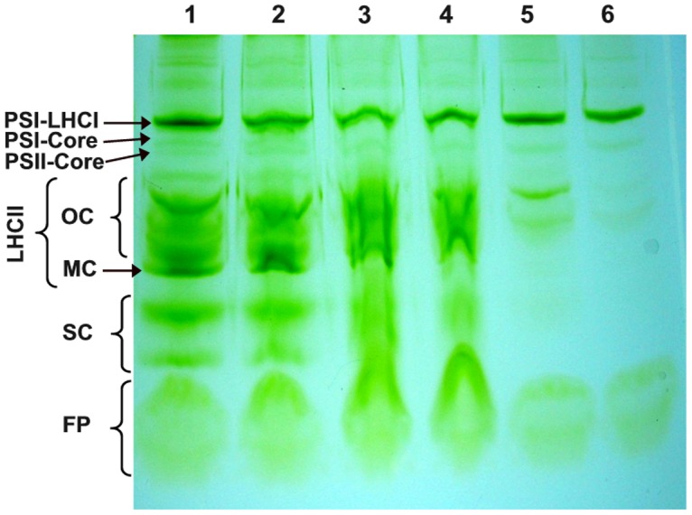
Lane 1: Control; lane 2: 1 mM Al3+ treated thylakoid membranes; lane 3: 2 mM Al3+ treated thylakoid membranes; lane 4: 3 mM Al3+ treated thylakoid membranes; lane 5: 4 mM Al3+ treated thylakoid membranes; lane 6: 5 mM Al3+ treated thylakoid membranes. Symbols on the left side indicate the following separated bands: PSI-LHCI, a number of large PSI complexes with attached LHCI antenna; PSI core, core protein complexes of PSI; PSII core, core protein complexes of PSII; LHCII OC, oligomeric LHCII complexes; LHCII MC, monomeric LHCII complexes; SC, small complexes; FP, free pigments. The electrophoretic patterns are representative for three independent experiments.