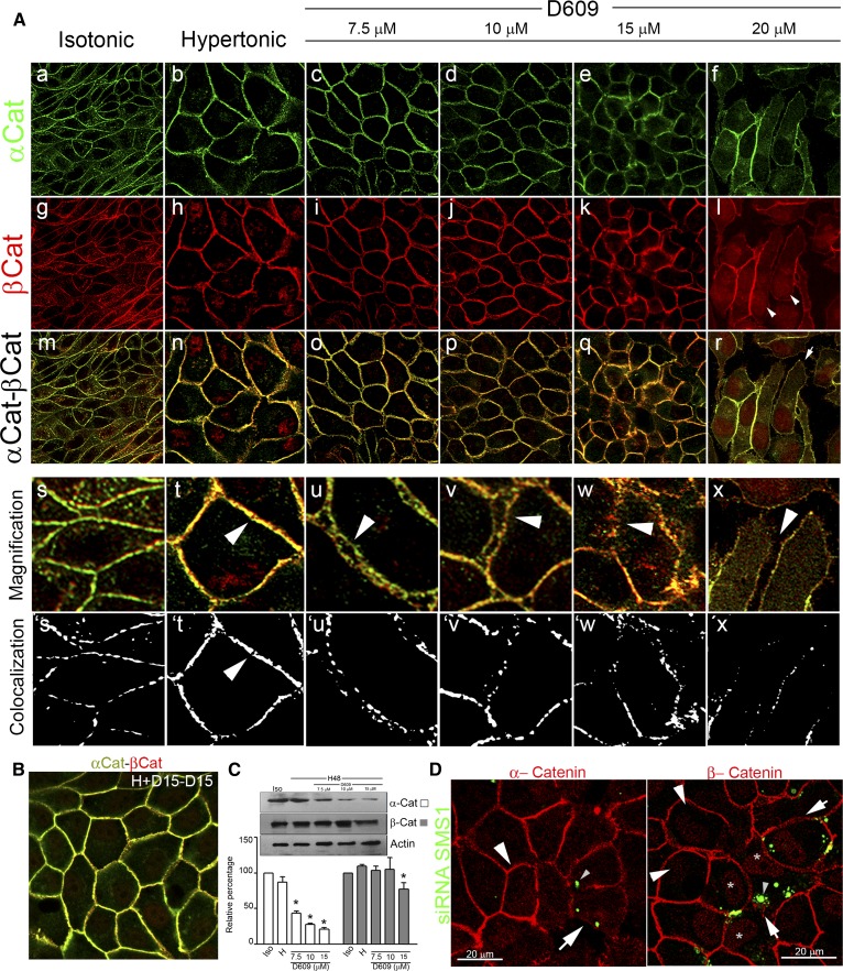
Fig. 5.
SMS inhibition and SMS1 knockdown impair AJ assembly: alteration of α- and β-catenin. A: Confocal immunofluorescence of the middle confocal plane of α-catenin (α-Cat) and β-catenin (β-Cat) distribution, under isotonic (a, g, respectively) or hypertonic conditions (b, h, respectively). Merged images (m, n). Confocal immunofluorescence of the middle confocal plane of α-catenin (c–f) and β-catenin distribution (i–l). Merged images of β- and α-catenin (o–r). Digital magnification (s–x). Arrowheads show the progressive alteration of AJ and cell-cell adhesion. From magnified images, segmentation processes were performed. Yellow pixels are represented as white pixels (Colocalization: s´, t´, u´, v´, w´, and x´). B: α-catenin (green) and β-catenin (red) were detected in wash cultured cells and reincubated without inhibitor. C: Effect of increasing concentrations of D609 on α-catenin (white bars) and β-catenin (gray bars) levels determined by Western blot analysis. Actin was used as loading control (representative image, n = 4, P < 0.05). D: Effect of SMS1 knockdown on α-catenin (red) and β-catenin (red) distribution. Transfected cells are seen as green dotted cells (small arrowhead).

