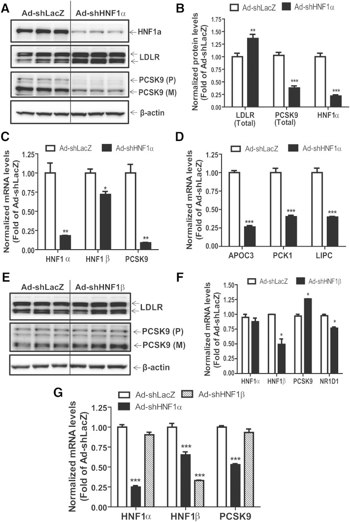
Fig. 2.
Knockdown of HNF1α results in elevated LDLR protein levels. HepG2 or mouse primary hepatocyte cells were seeded in 6-well or 24-well cell culture plates. After 24 h, cells were transduced with Ad-shLacZ (30 or 60 MOI), Ad-shHNF1α (30 MOI), or Ad-shHNF1β (60 MOI) adenoviruses. Fresh cell culture medium was replaced 6–8 h later and cells were incubated for a further 60–66 h. Total RNA and protein were collected from transduced cells for real-time PCR and Western blotting analyses. A, E: Western blotting with antibodies to LDLR, HNF1α, PCSK9, and β-actin was conducted by analyzing individual homogenates from Ad-shHNF1α- (A) or Ad-shHNF1β-transduced (E) HepG2 cells. B: Quantification of LDLR, PCSK9, and HNF1α from Ad-shLacZ- and Ad-shHNF1α-transduced HeG2 cells. C, D, F, G: Quantitative real-time PCR was conducted to determine the relative expression levels of mRNAs of interest from cultured HepG2 cells (C, D, F) or mouse primary hepatocytes (G). Data represent summarized result (mean ± SEM) of three independent replicates per treatment after normalization with GAPDH. *P < 0.05, **P < 0.01, and ***P < 0.001 compared with Ad-shLacZ transduced control cells which was set at 1.

