Fig. 4.
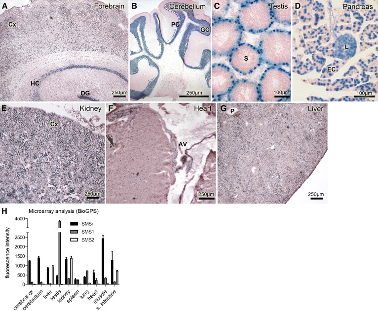
Mouse smsr is a ubiquitously expressed gene. β-galactosidase staining of cryo-sections from various SMSrD348E mouse tissues (A–G) correlates with smsr promoter activity. Neuronal regions of allo- and neocortical brain areas (A) and cerebellum (B) show prominent β-galactosidase staining mainly of nuclei and perinuclear areas of neurons. In testis, spermatogonia in the basal epithelium of the seminiferous tubules show similar nuclear β-galactosidase staining (C). In pancreas, β-galactosidase expression is displayed throughout the whole tissue, with a prominent staining of the exocrine portion and a somewhat weaker staining of the endocrine β-islets (D). In kidney, prominent β-galactosidase staining is displayed in the tubular system of the cortex (E). In heart, nuclei of all cardiomyocytes are β-galactosidase positive, especially those of the atrioventricular node (F). In liver, the distribution of β-galactosidase activity is diffuse with areas of more and others of less intensive staining that cannot be clearly associated to special regions of the hepatic lobules (G). AV, atrioventricular node; Cx, cortex; DG, dentate gyrus; EC, exocrine cells; GC, granular cell layer; HC, hippocampus; L, islets of Langerhans; P, portal field; PC, Purkinje cell layer; S, seminiferous tubule. H: Transcript expression profiles from microarray analysis of SMSr (Samd8), SMS2 (Sgms2), and SMS1 (Sgms1), modified according to BioGPS (47), data set Mouse MOE430 Gene Atlas (48). Microarray data were generated using an Affymetrix Mouse Genome 430 2.0 Array (GEO platform accession number GPL1261).

