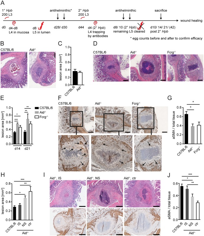
Fig 1. Antibody and Fcrg deficient mice display increased intestinal lesions and reduced accumulation of myofibroblasts.
Mice were challenge-infected with 200 infective Hpb larvae (scheme of infection and antihelminthic regime depicted in (A)), small intestines were harvested at day 10, 14 or 21 p.i.; tissue sections were stained by hematoxillin and eosin (H&E) or immunohistochemistry (IHC) and wide field microscopy images were analyzed using ImageJ. (B) Representative pictures of the largest H&E-stained cross sections of lesions at day 10 p.i. in C57BL/6 or Aid-/- mice; (C) Quantification of lesion area of largest cross sections (day 10 p.i.) for C57BL/6 or Aid-/- mice; (D) Representative pictures of the largest H&E-stained cross sections of lesions from C57BL/6, Aid-/- or Fcrg-/- mice at day 14 p.i.; (E) Quantification of lesion area of largest cross sections (day 14 or 21 p.i.) for C57BL/6, Aid-/- or Fcrg-/- mice; (F) Representative images of IHC staining for aSMA in largest cross sections of lesions in tissues from C57BL/6, Aid-/- or Fcrg-/- mice; arrows indicate aSMA+ areas with potentially contractile morphology. (G) Quantification of aSMA-stained area in lesions of C57BL/6, Aid-/- or Fcrg-/- mice; (H) Quantification of lesion area of largest cross sections (day 14 p.i.) for C57BL/6 or Aid-/- mice treated with immune serum from secondary infected (IS) or naïve (NS) C57BL/6 mice; (I) Representative pictures of the largest H&E-stained cross sections (top) or IHC staining for aSMA (bottom) of lesions at day 14 p.i. in C57BL/6 or Aid-/- mice treated with immune serum from secondary infected (IS) or naïve (NS) C57BL/6 mice; (J) Quantification of aSMA-stained area in lesions of C57BL/6, IS, NS or untreated (ctr) Aid-/- mice; All data are pooled from 2–3 independent experiments with 3–6 mice per group and presented as mean + SEM; Scale bars 200 μm.

