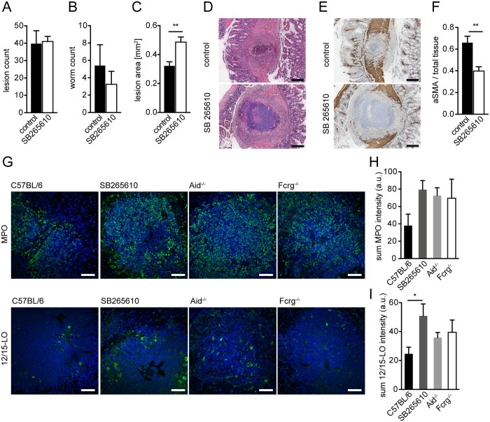
Fig 4. Inhibition of CXCR2 signaling leads to delayed MF accumulation and increased lesion size without affecting granulocyte recruitment in vivo.
C57BL/6 mice were treated or not with the CXCR2 antagonist SB265610 (3 mg/kg per oral gavage, once daily) during challenge infection with Hpb; small intestines were harvested for parasitology and histology at day 14 p.i. (A) Lesions were counted in small intestines of untreated (control) and SB265610 treated mice; (B) Adult worms were counted in opened small intestines of untreated (control) and SB265610 treated mice; (C) The area of the largest lesion cross sections was quantified in H&E-stained tissue sections from untreated (control) and SB265610 treated mice; (D) Representative H&E-stained cross sections of intestinal lesions in untreated (control) and SB265610 treated mice; Scale bars 200 μm. (E) Representative IHC-staining for aSMA in lesions of untreated (control) and SB265610 treated mice; Scale bars 200 μm. (F) Quantification of aSMA-stained area in lesions of untreated (control) and SB265610 treated mice was performed using ImageJ; (G) Small intestinal tissue sections from challenge-infected untreated C57BL/6 (control) and SB265610 treated C57BL/6 mice or Aid-/- or Fcrg-/- mice were IF-stained for the neutrophil marker MPO (upper panel, green) or the eosinophil marker 12/15-lipoxygenase (12/15LO) (lower panel, green), followed by counterstaining with DAPI (blue); Scale bars 50 μm. (H) Total Fluorescence intensity of MPO was quantified using CellProfiler; (I) Total Fluorescence intensity of 12/15LO was quantified using CellProfiler; All data are pooled from at least 2 independent experiments with 3–6 mice per group and presented as mean + SEM.

