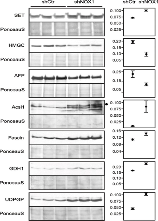
Fig 4. Western blot validation of NOX1 regulated proteins.
The major components of the 2DE spots which contained complex proteins mixtures were subjected to validation through Western blot analysis. Seven of the proteins were confirmed to be differentially expressed in control vs. NOX1 reduced HepG2 cells. Plotted are the mean and SEM of WB intensities normalized to the total protein content of the sample determined from the PonceauS staining. SET—protein SET, HMGC—HMG-CoA-synthase, AFP—Alpha-fetoprotein, Acsl1—Long chain fatty acid-CoA ligase, GDH1—Glutamate dehydrogenase 1, UDPGP—UTP-glucose-1-phosphate uridylyltransferase. Statistical results are presented in Table 1.

