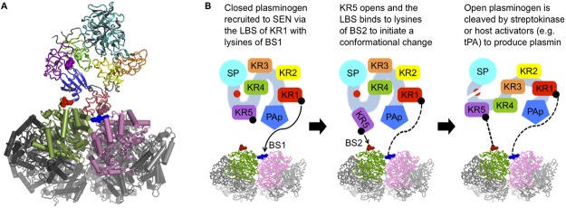
Fig 9. Molecular docking results and mechanism of plasminogen binding by SEN.

(A) Human plasminogen KR1 docked at the SEN minor interface. Plasminogen domains are colored as follows: pan-apple (PAp) domain blue; KR1 red; KR2 yellow; KR3 orange; KR4 green; KR5 purple; serine protease (SP) domain cyan. Linear three-residue patches on KR1 and KR5 implicated in lysine binding are shown as spheres. One SEN dimer is colored green and pink, respectively, with the remaining octamer colored in shades of grey. One occurrence of SEN BS1 and BS2 is shown as blue and red spheres, respectively. (B) Proposed model for the binding of plasminogen by SEN involving interactions between BS1 and KR1, and BS2 and KR5. The color scheme follows (A). Figure adapted from Law et al. [22].
