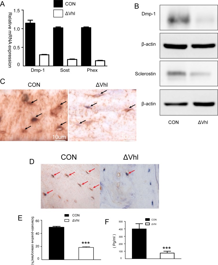
Fig 2. The role of HIFα pathway in the differentiation of osteocytes.
(A) Quantitative PCR analysis of the differentiation markers of osteocytes. (B) Western blot analysis of DMP-1 and sclerostin proteins in the tibia of 2-month-old CON and ΔVHL mice. (C) Immunohistochemical analysis of DMP-1. Compared to the findings from CON, DMP-1 expression in the conditional ΔVHL osteocytes is dramatically reduced (arrows). (D) Immunolocalization of sclerostin in transverse sections of the mid-femoral diaphyses of 6-week-old mice. (E) The percentage of sclerostin-positive osteocytes in the mid-femoral diaphyses of the CON and ΔVHL mice (n = 6 mice for each genotype *** p < 0.001). (F) Serum levels of sclerostin in 3-month-old CON and ΔVHL mice (n = 9 mice for each genotype *** p < 0.001).

