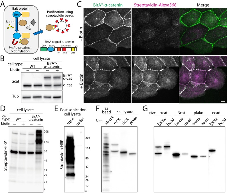
Fig 1. Characterization of promiscuous BirA (BirA*)-tagged α-catenin expressing cells.
(A) Illustration of in situ proximal biotinylation and subsequent purification of biotinylated proteins. A stable cell line expressing BirA*-α-catenin grown in biotin containing media with subsequent biotinylated proteins purified with magnetic streptavidin-beads. Schematic of a BirA*-α-catenin construct. The BirA* is flanked by GFP and α-catenin. (B) Western blots of the wildtype (WT) and BirA*- α-catenin expressing MDCK stable cell lines. The cell lysates were analyzed using anti-α-catenin (top) and anti-tubulin antibodies (bottom). (C) The BirA*-α-catenin expressing cells were treated with biotin for 24 hours, then analyzed for the localization of BirA*-α-catenin and biotinylated proteins using AlexaFluor-568 labeled streptavidin. BirA*-α-catenin localized to cell-cell contacts, and in the presence of biotin in the media, streptavidin-specific labeling localized to cell-cell contacts, demonstrating the proximal biotinylation by BirA*-α-catenin. Scale bar 20 μm. (D) Detection of biotinylated proteins in BirA*-α-catenin expressing cells. The wildtype (WT) or BirA*-α-catenin expressing stable cell lines were cultured with or without biotin for 24 hours. The cell lysates were analyzed using western blots with streptavidin-HRP. (E) Most biotinylated proteins were in the soluble pool of cell lysates. Post-sonication cell lysates were centrifuged at 16,000g for 20 minutes, and the supernatant and pellet were analyzed using Western blot with streptavidin-HRP. (F) A streptavidin-bead purified sample from cells plated on a P150 dish was analyzed using Western blot with streptavidin-HRP. In adjacent lanes, cell lysate was analyzed using catenin antibodies. The molecular weights of major bands in the purified sample correspond to that of catenins. (G) Cell lysates and purified samples (bead) from cells plated on a P150 dish were analyzed with catenins and E-cadherin antibodies. The catenins were present in the bead fractions but E-cadherin was not.

