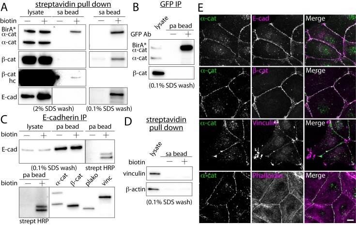
Fig 2. In situ proximal biotinylation by promiscuous BirA*-α-catenin.
(A) Western blots of purified proteins from BirA*-α-catenin expressing cells using streptavidin-conjugated beads. E-cadherin (E-cad), β-catenin (β-cat), vinculin and β-actin antibodies. Both 2% and 0.1% SDS wash conditions shown. The image contrast of β-catenin blot was enhanced to show the presence of β-catenin in the bead fraction (β-cat hc). (B) Immuno-precipitation of GFP-tagged BirA*-α-catenin proteins using an anti-GFP antibody. The immuno-precipitaed samples were analyzed using Western blot with α-catenin and β-catenin antibodies. (C) Immuno-precipitation of E-cadherin from BirA*-α-catenin expressing cells using an E-cadherin antibody. From immuno-precipitaed proteins, two readily identifiable bands in the streptavidin blot had identical molecular weights as that of β-catenin and plakoglobin. (D) Western blots of strepavidin purified proteins using vinculin and β-actin antibodies. Vinculin and β-actin were not detected in the streptavidin purified protein pool. (E) Co-localization analysis of α-catenin and other cell-cell adhesion and cytoskeletal proteins. While E-cadherin, β-catenin, vinculin and actin filaments co-localize with α-catenin, only E-cadherin and β-catenin are biotinylated by BirA*-α-catenin. Scale bar 10 μm.

