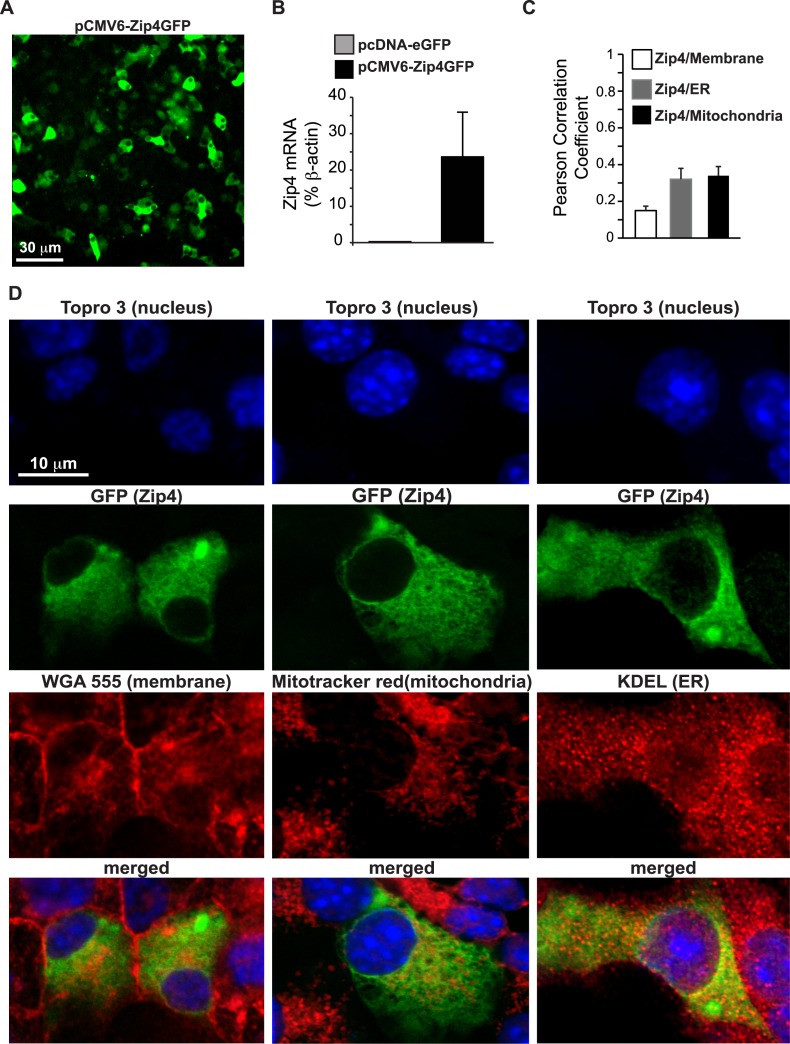
Fig 2. Localization of Zip4 in MIN6 cells.
A. Field of view of MIN6 cells transfected with the pCMV6-Zip4GFP plasmid. B. Expression of Zip4 was measured by qPCR performed on MIN6 cells transfected with pCMV6-Zip4GFP plasmid C. Co-localization was estimated with the Pearson correlation coefficient analysis calculated from confocal images obtained from the FITC and Cy3 channel. D. Confocal images of pCMV6-Zip4GFP transfected MIN6 cells after staining of the nucleus with TOPRO-3, plasma membranes with WGA Alexa Fluor 555, mitochondria with MitoTracker Red CMXRos or the endoplasmic reticulum (ER) with immunostaining of KDEL.

