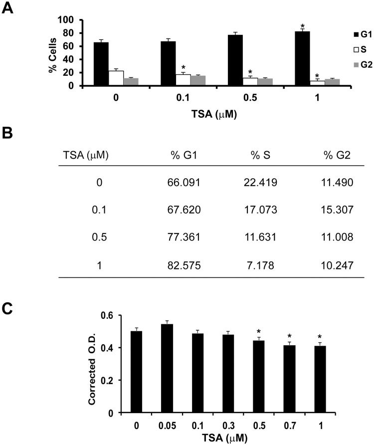
Fig 1. TSA induces RPE cell cycle arrest by inhibiting cell proliferation.
Cell cycle analysis of RPE cells treated with 0–1 μM TSA for 24 h, fixed with ice-cold 70% ethanol and stained with propidium iodide by an EPICS XL-MCL flow cytometer. (A) With increasing doses of TSA, significantly more RPE cells were in the G1 phase and fewer in the S phase than untreated cells. (*: t test p<0.01) (B) Percentage of cells in each phase of the cell cycle after TSA treatment. (C) PrestoBlue assay was performed on RPE cells treated with 0, 0.05, 0.1, 0.3, 0.5, 0.7 or 1 μM TSA to determine the toxicity of TSA on RPE cells. The lowest number of viable RPE cells was seen when cells were treated with 1 μM TSA for 24 h (82.0% viable cells). (*: t test p< 0.0001).

