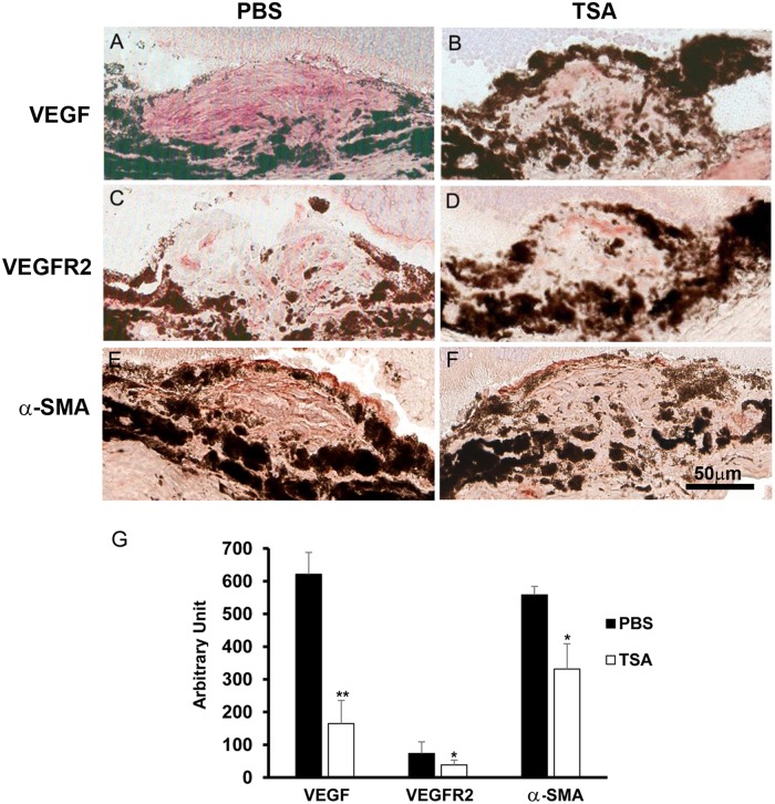
Fig 14. TSA reduces VEGF, VEGFR2 and α-SMA in mouse CNV lesions.
Immunohistochemical staining was performed on murine retinal cryostat sections in CNV lesions day 7 post-laser for (A-B) VEGF, (C-D) VEGFR2 and (E-F) α-SMA. Figures on the left panel (A, C and E) are from PBS control mice, and figures on the right panel (B, D and F) are from TSA-treated mice. TSA reduced the amount of cells stained positively for (A) VEGF, (C) VEGFR2 and (E) α-SMA, when compared to PBS controls (B, D and F). (G) Quantification of positively stained area for each protein normalized by the size of the CNV lesions. (*: t test p<0.05; **: t test p<0.01; n = 4/group; bar = 50 μm).

