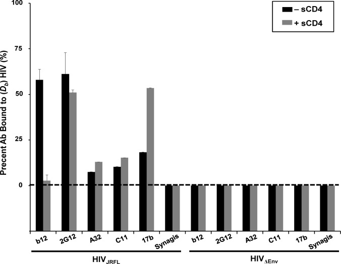
Fig 1. Anti-envelope mAb binding to HIVJRFL virions in solution measured by Fluorescence Correlation Spectroscopy (FCS).
HIVJRFL virions (10μg/ml p24 equivalent concentrations) were allowed to interact in solution with Alexa Fluor 647-conjugated test Mabs b12, 2G12, A32, C11, and 17b, or negative control antibody Synagis in a 100μL volume at 4.5–6.6μg/ml final concentrations (see Methods). The relative fraction of Mabs that adopts a slower diffusion coefficient (D b~8 μm2 /sec) as a result of virion binding is indicated by black bars. The percentage of Mab binding to virions treated with 100μg/ml sCD4 is shown in grey bars. As a control for nonspecific binding, Mabs were tested for interactions with particles not expressing HIV Env (HIVΔENV). All experiments were repeated at least four times, and average values are shown. Error bars indicate standard deviation.

