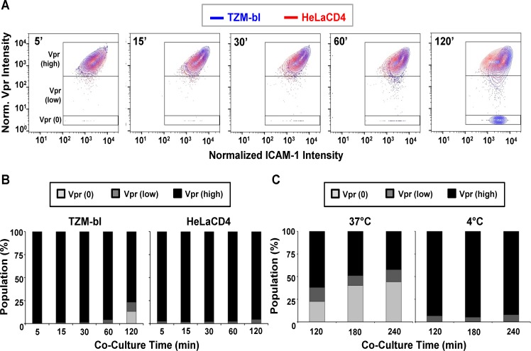
Fig 3. Changes in SNAP-ICAM-1 and CLIP-Vpr signals on HIVJRFL pseudoviruses attached to TZM-bl or HeLa-CD4 target cells.
(A) To characterize changes in HIVJRFL during interactions with target cells, ROIs comprising cell-bound virus particles were selected and used to extract SNAP-ICAM-1 and CLIP-Vpr intensity signals. These data were then normalized in order to generate contour plots (see Methods) of HIVJRFL signals on TZM-bl (blue) or HeLa-CD4 (red) target cells at the indicated times. Three Boolean gates were defined based on Vpr content at the 5 minute co-culture time point with TZM-bl cells: Vpr (0), corresponding to data points with no Vpr signal (bottom box); Vpr (low) corresponding to data in the bottom 5th percentile of signal intensity (middle box); and Vpr (high) corresponding to the top 95th percentile of the data set (top box). A minimum of 1800 ROI per condition are shown. (B) Based on the gating strategy described in (A), the Vpr (0) (light grey), Vpr (low) (dark grey), and Vpr (high) (black) subpopulations were quantified as percentages of the entire bound virus population on TZM-bl cells (left) or HeLa-CD4 cells (right) after the indicated times of attachment. (C) The same three subpopulations were quantified on TZM-bl cells after extended attachment periods (120–240 minutes) at the fusion permissive temperature 37°C (left) or at 4°C (right) where membrane mixing is arrested.

