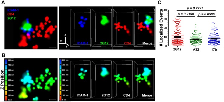
Fig 7. Superresolution imaging of Gp120 epitope exposure on TZM-bl-bound HIVJRFL virions.
HIVJRFL virions were co-cultured with TZM-bl cells for 30 minutes and fixed. Gp120 epitope exposure was probed with Alexa 488-conjugated Mabs. (A) Top: A representative lateral dSTORM projection image of a virion bound to a TZM-bl cell. Cell surface CD4 is stained with Alexa 647-conjugated, anti-CD4 antibody OKT4 (red). The virion surface is marked by ICAM-1 tagged with membrane-impermeable SNAP-Alexa 546 (Blue); gp120 is stained by Alexa 488-conjugated Mab 2G12 (green). Scale bar = 0.1μm. Bottom: Image matches the one above, but with Z position scaling. The Z imaging range is divided into 100 nm sections; each is color coded as shown. Thus, signals with the same color fall within the same section (i.e. within 100nm). (B) Axial view of the dSTORM image in A, with the color channels separated as well as merged together. The Z line in the color channel panel is pointing upward away from the cell. The bottom images match the ones above, but with Z position scaling as in (A). Note that the ICAM signals are comprised within no more than two adjacent 100nm sections. (C) The number of localized events generated by Mabs 2G12, A32, or 17b in at least 110 superresolution ROIs containing bound HIVJRFL virions is shown (see Methods). Black bars indicate the geometric mean and standard errors. The two-tailed Mann-Whitney test was used to perform pairwise comparisons of localized events measured with each Mab.

