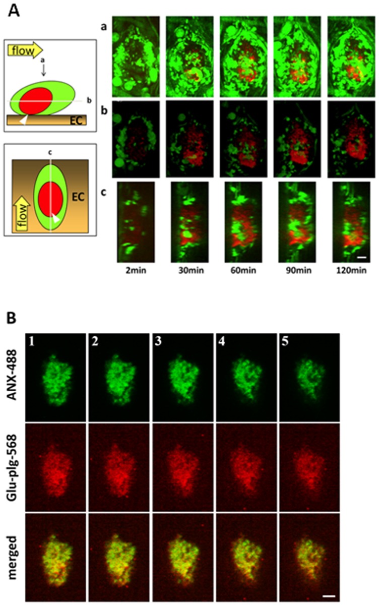
Fig 2. Spatiotemporal distribution of Glu-plg-568 within the microthrombus.
(A) Localization of Glu-plg-568 in a microthrombus in a GFP mouse. a) images of the whole thrombus from the luminal side. b) horizontal plane (X-Y) images at the point where the area of Glu-plg-568 was the largest. c) perpendicular plane (Y-Z) images at the point of the longest Y- axis. Images were taken at 2, 30, 60, 90, and 120 min after laser injury. The schematic depiction shows the horizontal and vertical planes of a laser-induced thrombus containing Glu-plg-568 (red) and GFP platelet (green) on the injured endothelial cells (EC). Glu-plg-568 accumulated in the center of the microthrombus in a time-dependent manner. The arrow shows the direction of blood flow. Scale bar, 10 μm. (B) Co-localization of the PS (ANX-488) and Glu-plg-568 within the microthrombus. ANX-488 and Glu-plg-568 were injected into the tail vein of WT mice before laser injury. Images were collected 60 minutes after the mesentery injury. Five optical sections (1–5) were selected at 2-μm intervals from the vessel wall (1) to the luminal surface of a thrombus (5) to examine the spatial distribution of PS-exposing platelets and Glu-plg. Localization of Glu-plg-568 within the microthrombus paralleled the localization of ANX-488 bound to the surface of PS-expressing platelets (n = 3 thrombi from three mice). Scale bar, 10 μm.

