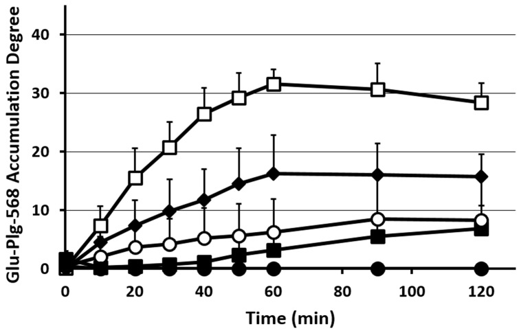
Fig 3. Changes in Glu-plg-568 accumulation in microthrombi after laser injury.
The kinetics of Glu-plg-568 accumulation are shown as an increase in the integrated fluorescence intensity of Glu-plg-568 per corresponding thrombus area in the same optical section 60 minutes after laser-induced thrombus formation. Reagents were EACA (closed squares, N = 5, 5 thrombi from 3 mice, p < 0.001), CPB (closed diamond, N = 5, 5 thrombi from 5 mice, p < 0.005) and aprotinin (open circles, N = 4, 4 thrombi from 2 mice, p < 0.001). Control experiments are shown as open squares (N = 5, 5 thrombi from 5 mice) and mini-plg-568 as closed circles (N = 4, 4 thrombi from 4 mice, p < 0.001). This assay was analyzed with repeated measures ANOVA. Each point represents the mean ±SD; N = number of thrombi.

