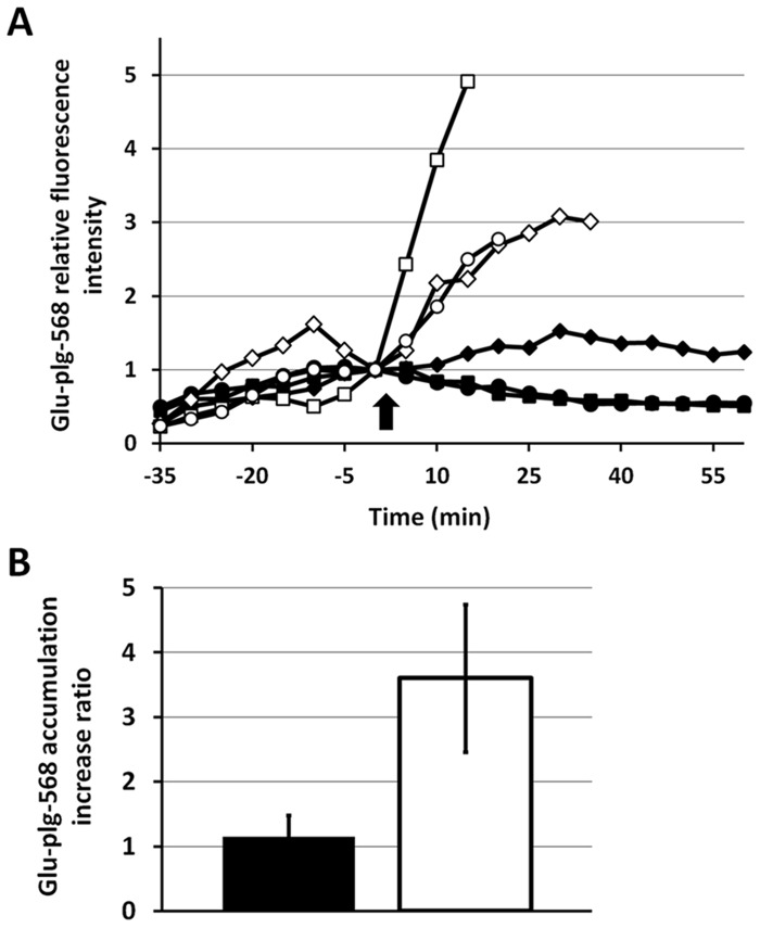
Fig 7. Changes in Glu-plg-568 relative fluorescence intensity induced by exogenous tPA administration.
(A) Glu-plg-568 was administered to GFP-mice 10 minutes before each laser injury and 40 minutes after either human tPA (white: square, diamond, circle; three individual experiments) or an equivalent volume of 0.9% NaCl (black: square, diamond, circle; three individual experiments) was infused. Thrombi images were collected every 5 minutes after the laser-evoked endothelial injury, starting 2 minutes before and then every 5 minutes up to 60 minutes after either tPA or saline infusion. Each fluorescence intensity was normalized to the value obtained 2 minutes before either tPA or saline administration. tPA or saline was injected at time 0 (arrow). (B) Shown are the Glu-plg-568 accumulation increase ratios, which were expressed as the average values of the maximum increases in Glu-plg-568 relative fluorescence intensity after administration of either tPA (mean ± SD, n = 3, white column) or saline (mean ± SD, n = 3, black column). This assay was analyzed with a t-test for independent samples (P < 0.05).

