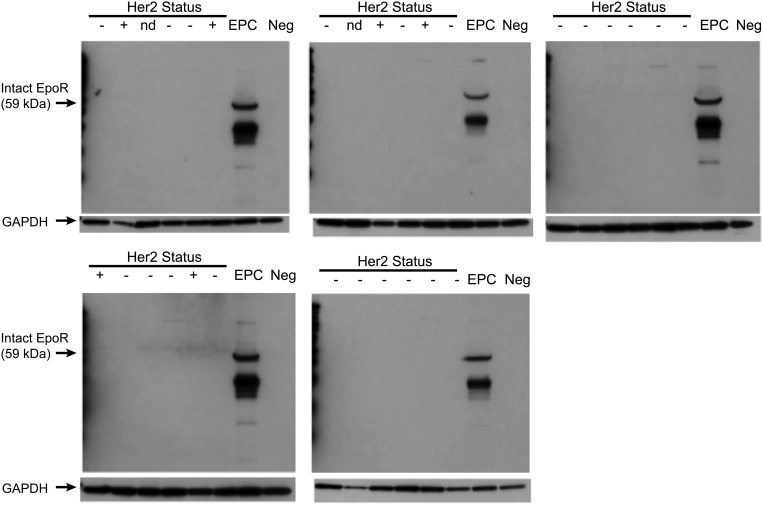
Fig 8. Analysis of EpoR protein expression in breast tumor tissues.
Lysates prepared from an aliquot of tumor tissue from 30 patients from the cohort of breast tumor tissues that were analyzed by flow cytometry. Western blot analysis of EpoR expression was performed using the EpoR specific monoclonal A82. Erythroid progenitors (EPC) obtained following 8 days of differentiation in vitro from human bone marrow isolates cultured in the presence of IL-6, SCF, and rHuEpo were used as positive controls. GAPDH was used as a loading control.

