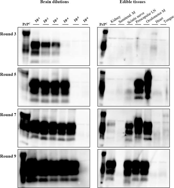
Fig 3. Converting activity in the inocula from 1216B sheep analysed by vPMCA.
Western blot analysis of the amplified products was carried out after the 3rd, 5th, 7th and 9th rounds, as indicated. The results obtained with the brain dilution curve are reported in left panels, those from visceral tissue inocula are in right panels. Blots were probed with SAF84 primary antibody.

