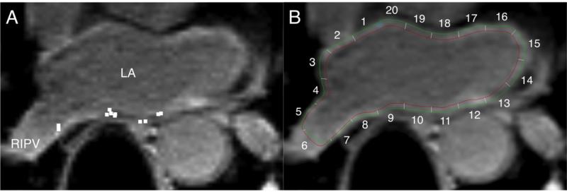
Figure 2. Registration of EAM data to post-ablation LGE-MRI.
A: Location data of ablation sites on EAM (white square dots) were registered to the post-ablation MRI by using custom software. B: The LA endocardial and epicardial contours are drawn onto the axial image at the same level as Panel A. LA sectors on axial image planes corresponding to ablated sites were identified. In this example, sectors 7, 9, 10, 11 were identified as ablated sectors.

