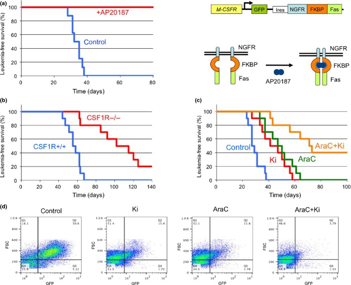
Figure 6.
Cure of mixed lineage leukemia (MLL)-AF10-induced acute myeloid leukemia (AML) by ablation of cells expressing high levels of macrophage colony-stimulating factor (M-CSF) receptor (CSF-1Rhigh). (a) Bone marrow (BM) cells from the transgenic (CSF-1R-EGFP-NGFR/FKBP1A/TNFRSF6) mice were infected with MSCV-MLL-AF10-ires-GFP and were transplanted into lethally irradiated C57BL/6 mice to induce AML. BM cells (1 × 10⁴) of primary AML mice were transplanted into sublethally irradiated C57BL/6 mice. Administration of AP20187 or solvent (control) to the secondary AML mice was started by i.v. injection 3 weeks after transplantation. Leukemia-free survivals of the untreated (n = 8) and AP20187-treated (n = 8) secondary transplanted mice were investigated (P < 0.001). Right panel shows the structure of genes for the Csf1r promoter, EGFP, the NGFR–FKBP–Fas suicide construct, and activation of NGFR–FKBP–Fas. Note that in the transgenic mice, conditional ablation of cells expressing high levels of CSF-1R can be induced by exposure to the AP20187 dimerizer. (b) Fetal liver cells of E16.5 Csf1r+/+ and Csf1r−/− mice littermate embryos were infected with MLL-AF10-ires-GFP and transplanted into irradiated mice. The leukemia-free survivals of the mice were analyzed (n = 10, P < 0.001). (c,d). BM cells (105) from AML mice with MLL-AF10 were transplanted into non-irradiated mice. Mice were treated with vehicle, Ki20227 (Ki), Ara-C, or Ki plus Ara-C (AraC+Ki). Leukemia-free survivals were analyzed (c) (n = 10, P < 0.01 [control vs Ki; AraC vs AraC+Ki]). Peripheral blood cells were prepared 21 days after transplantation and analyzed for expression of GFP (d).

